 +91 85238 33348
 kumarpre@hotmail.com
A Camera Inside the Heart Artery? OCT vs IVUS for Complex Angioplasty and Left Main Stenting
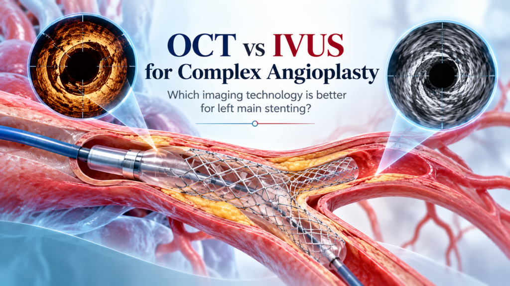
An angiogram shows your heart arteries as a shadow. Useful, but a shadow cannot tell a doctor exactly how thick the plaque is, or whether a stent is pressed fully against the artery wall. For that, cardiologists now send a tiny camera inside the artery itself.
Two technologies do this: IVUS and OCT. A 2026 trial compared them in one of the most demanding angioplasties there is.
Meet the two cameras
| IVUS | OCT | |
|---|---|---|
| Uses | Sound waves (ultrasound) | Light (near-infrared) |
| Think of it as | A sonar that sees deep into the artery wall | A high-definition camera with very fine detail of the inner surface |
| Strength | Sees the full thickness of large arteries; no extra dye needed to take pictures | Sharper images of stent struts, calcium and small tears |
| Trade-off | Lower image resolution | Needs a flush of contrast dye; sees less deeply |
Why the left main artery is special
The left main is the short trunk that supplies most of the heart muscle before it splits into two major branches. A blockage right at that fork, called a distal left main bifurcation, is among the highest-stakes places to put a stent. Sizing and positioning have to be exact.
Because the left main is a large vessel, IVUS has long been the preferred imaging tool there. Whether OCT could do the job equally well was an open question.
What the 2026 trial found
The ISOLEDS trial, presented at the European Society of Cardiology Congress 2026, randomly assigned 664 patients with true distal left main bifurcation disease at 24 hospitals in China to OCT-guided or IVUS-guided stenting.
“Failure” meant cardiac death, a heart attack related to the treated segment, or a repeat procedure on it. OCT was noninferior to IVUS: it performed at least as well. The gap between the two numbers was not large enough to say OCT is better.
What happens during an imaging-guided angioplasty
1Before the stent: the camera measures the artery and the plaque, so the right stent size and length are chosen.
2Preparing the blockage: if heavy calcium is seen, it can be treated before the stent goes in.
3After the stent: the camera checks that the stent is fully expanded and sitting flush against the wall, with no tears at the edges.
4Fine-tuning: anything imperfect is corrected before the procedure ends.
For the patient, it is the same procedure through the same small puncture, usually with a few extra minutes for imaging.
Why an angiogram alone is sometimes not enough
A coronary angiogram fills the artery with dye and takes an X-ray. What the doctor sees is the outline of the channel the blood flows through. It does not show the artery wall itself. Three things are therefore hard to judge from an angiogram:
- The true size of the artery. Plaque can be spread evenly around the wall, making a diseased artery look simply “small”.
- What the plaque is made of. Soft fatty plaque and hard calcium behave very differently when a balloon is inflated.
- Whether a stent is fully open. A stent can look acceptable on X-ray and still be under-expanded or not touching the wall in places.
Under-expanded stents are one of the main reasons a stent narrows again or clots. Imaging from inside the artery is how these problems are found and fixed during the procedure, not months later.
What the doctor is looking for on the images
| Finding | What it means | What is done about it |
|---|---|---|
| Stent under-expansion | The stent has not opened to its intended size | Further balloon inflation at higher pressure |
| Malapposition | Part of the stent is not in contact with the artery wall | A larger balloon to press it into place |
| Edge dissection | A small tear in the artery at the end of the stent | Observation if minor, or an additional stent |
| Heavy calcium | The plaque is too hard for a stent to expand properly | Calcium is modified first with specialised balloons or devices |
Left main disease: stent or bypass?
Because the left main supplies such a large part of the heart, a significant blockage there is always taken seriously. The two established treatments are bypass surgery and angioplasty with stenting. Which is better for an individual depends on how many other arteries are diseased, how complex the blockages are, whether the patient has diabetes, heart pumping function and fitness for surgery. This decision is ideally made jointly by a cardiologist and a cardiac surgeon. When stenting is chosen, imaging guidance is strongly favoured because the margin for error is small.
Why a bifurcation makes stenting harder
At a fork, plaque often extends into both branches. The operator must keep both branches open, decide whether one stent or two are needed, and make sure the metal overlaps cleanly where the branches meet. Each of those steps is easier to get right when the artery can be seen from the inside.
Which patients may suit which tool?
- IVUS is often preferred when kidney function is reduced, because it does not need extra dye, and when the blockage sits right at the very mouth of the left main artery, a spot that is difficult to clear of blood for OCT.
- OCT is often preferred when very fine detail is needed, such as assessing calcium thickness, checking stent struts or working out why an older stent has failed.
- In many patients either tool works well, which is what the 2026 trial supports for distal left main bifurcation disease.
Are there any risks from the imaging itself?
Intravascular imaging is considered safe and complications from the imaging catheter are uncommon. It adds a little time to the procedure, and OCT adds some contrast dye. Your cardiologist weighs these small additions against the benefit of a better-placed stent.
So, which one is better?
Keep in mind that this was a single trial in one country with one year of follow-up, and both doctors and patients knew which tool was used.
The bottom line
For complex left main stenting, the question is shifting from “which camera?” to “is a camera being used, and by someone experienced in reading it?”
Frequently asked questions
Is the imaging catheter painful?
No. It travels over the same thin wire used for the angioplasty and you do not feel it inside the artery.
What is the difference between OCT and IVUS in simple terms?
IVUS uses sound waves and sees deeper into the artery wall. OCT uses light and gives sharper detail of the inner surface and the stent. Both produce cross-section pictures of the artery from the inside.
Which is better for left main stenting, OCT or IVUS?
In the 2026 ISOLEDS trial, OCT guidance was noninferior to IVUS guidance at 12 months. Neither was shown to be superior. The choice depends on anatomy, kidney function and operator experience.
Does every angioplasty need OCT or IVUS?
Not necessarily. It is most valuable in complex cases: left main disease, bifurcations, long or heavily calcified blockages and failed previous stents.
Does imaging make the procedure much longer?
It usually adds a few minutes. In complex cases this time is spent making sure the stent result is as good as it can be.
Is OCT safe if I have kidney problems?
OCT needs additional contrast dye, so in patients with reduced kidney function IVUS is often chosen. Tell your cardiologist about any kidney condition before the procedure.
Can I ask whether imaging will be used in my angioplasty?
Yes. It is reasonable to ask whether imaging guidance is planned, which type and what it will be used to check.

This article is for general patient education and is not a substitute for a consultation. Treatment decisions depend on your individual reports, anatomy and overall health, and should be made with your cardiologist.